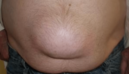
Surgery of ventral hernias (umbilical and epigastric) and small incisional hernias.
Umbilical hernia and epigastric hernia (located above the navel) are a hole in the abdominal wall. We can assimilate to these hernias, the small incisional hernia due to a weakness on the abdominal scar, either next to a classic scar of opening of the belly, or next to a small scar of endoscopic surgery (trocar orifice). The diagnosis can be made by the mere presence of a bulge that can increase in volume during efforts. In other situations, it is the thinning of the skin, and the appearance of skin disorders (bluish appearance, etc.) that can attract attention. Discomfort or pain, the unfolding of the umbilicus (photo), the presence of intestine in the hernia are usually indications for surgery.
Bulky umbilical hernia - Umbilical hernia


Epigastric hernia
The hernia can be responsible for discomfort, even pain, especially during physical activity. The main complication is hernial strangulation with the incarceration of fatty tissue or an intestinal loop, the hernia then becomes very painful, vomiting is frequent and the surgical treatment becomes very urgent. If the treatment is carried out less than 6 hours from the episode of strangulation, it is often possible to reintegrate the intestine trapped in the hernia and to repair the wall correctly, provided that the practitioner who operates on you in emergency is trained in the technique of parietal surgery.
After 6 hours of strangulation, the part of the intestine stuck in the hernia necroses and must be removed, the surgical intervention is then more delicate for the patient who is not prepared and who sometimes takes anticoagulant type treatments . In this emergency context, the priority for the surgeon is to treat the intestinal obstruction and to perform the ablation and then the intestinal suture. In this context, a surgeon, even partially specialized in parietal surgery, will not always be able to carry out a good repair of the wall, because the use of a prosthetic reinforcement will often be contraindicated. The risk of the hernia coming back (recurrence) is then very high.
It is for these reasons that the presence of a ventral hernia should lead to a surgical consultation in search of a possible surgical indication to be performed in a scheduled surgery.
An ultrasound is sometimes useful, it must be requested by the specialist. The clinical examination performed by the surgeon is usually sufficient for the diagnosis. Of course, in the presence of a small old and stable hernia and in the absence of any sign of seriousness, a simple monitoring can be decided.
In our daily practice we implement our concept of minimally invasive surgery for umbilical and epigastric hernias and small incisional harnias by aesthetic mini incision.

The use of a parietal reinforcement prosthesis is practically systematic. However, for the smallest hernias whose size does not exceed a few millimeters, a simple suture can be proposed. In the vast majority of cases, the treatment can be carried out in a minimally invasive way, preferably during a hospital stay of a few hours.
Left picture: Example of ventral parietal reinforcement prosthesis.
Specificity of postoperative abdominal wall weakness problems related to laparoscopic surgery:
Since the generalization of laparoscopic surgery techniques to perform removals of the gallbladder, the appendix of the colon of the prostate, etc., the surgeon most often no longer makes large incisions, but even the small scars of the openings to introduce trocars , and especially at the umbilical level can be the site of small incisional hernia which can evolve with a risk of intestinal obstruction by trapping the small intestine. They then require specific care with a treatment that must be adapted and sufficient from the outset with the use of elaborate techniques. Insufficiently treated (without prosthesis for example) these “small” incisional hernias can return (it is the recurrence), and require re-interventions much more complicated and sometimes dangerous.
How to operate a ventral hernia and why would we use a prosthesis

The use of a parietal reinforcement prosthesis is practically systematic for the treatment of ventral hernias, except for the smallest hernias whose size does not exceed a few millimeters, or a simple suture can be proposed. The prosthetic material can be inserted either openly (small aesthetic incision in front of the hernia) or by laparoscopic surgery.
The use of laparoscopic surgery in our experience is limited to rare cases, because this technique can lead to specific, rare, but sometimes serious intraoperative complications (intestinal wounds, vascular injuries, etc.), and the placement of prosthetic material in intraperitoneal position can also create postoperative adhesions and lead to late complications (intestinal obstruction, migration of prosthetic material, etc.).
New laparoscopic or even robot-assisted techniques are currently being developed, they are already very interesting today for certain forms of incisional hernias. But they are longer, more invasive compared to the techniques with mini incision that I use for the most common cases.

In the vast majority of cases, the prosthesis is therefore applied within the abdominal wall, through a small aesthetic incision under “light” general anesthesia (without endotracheal intubation or curare), during a hospital stay of a few hours. The patient can take his shower the day after the intervention, without specific postoperative care.
Large postoperative incisional hernia: Conditions of occurrence, diagnosis, when and how to operate, the contribution of the reinforcement prosthesis placed in the abdominal wall.
The surgical treatment of large post-operative incisional harnias is one of my specialties.
Incisional hernia results from the release of an abdominal scar after a large opening (laparotomy).
Incisional hernia is more frequent in the presence of risk factors (congenital weakness of the aponeuroses, obesity, postoperative infection, etc.).
Incisional hernia is manifested by a bulge on a scar.
At first, the bulge spontaneously disapears to the supine position. The incisional hernia contains intra abdominal fat, or bowel.
The evolution of this incisional hernia is usually done by an increase in volume of the bulge which is no longer reduced, and the appearance of pain.A very sharp pain accompanied by nausea or vomiting can be a sign of intestinal obstruction, requiring treatment in extreme emergency.
Any incisional hernia, even a beginner, requires a specialist consultation, in order to carry out an assessment, to specify whether surgery is necessary, or whether simple monitoring can be recommended. (small old incisional hernia without discomfort, not progressive…). During the consultation and if necessary the type of intervention to be proposed will be discussed.
Many patient-related factors (Medical history, morphology, etc.), or related to the incisional hernia itself (volume of the incisional hernia, pain, etc.), often with the result of a CT scan, will guide towards surgical treatment using practically always a reinforcement prosthesis, it is a mesh or a tulle sometimes incorrectly called “plate”, indeed in fact this reinforcement is very flexible and malleable. The reinforcement will most often be placed openly, by incising all or part of the initial scar.
I must specify that I am most often in favor of the open route (incision on the scar), in order to be able to put the prosthetic material within the abdominal wall itself and to isolate it from the intra abdominal viscera. Indeed positionning the prosthesis in the abdominal cavity by endoscopic surgery, can sometimes be complicated by harmful adhesions between the prosthesis and the intra-abdominal viscera. with the risk of migration of the prosthesis and with the risk of recurrence even years after the repair of the incisional hernia. With the risk of strangulation or peritonitis with the need for an often complex reoperation, with a sometimes severe prognosis. Our participation in most of the international congresses concerning this theme shows in fact a limitation of the use of endoscopic surgery compared to the open route for the incisional hernias that we usually treat. Ongoing studies will clarify the role of new robot-assisted techniques, some incisional hernias can be operated now with a robot, but only some spécific cases. Our personal practice for the moment and for the more common cases is to perfect conventional techniques with small or medium incisions, and to put large prostheses during short-term hospitalization.

Si la hernie de l'aine ou la hernie ventrale (ombilicale, épigastrique) sont dues à une faiblesse congénitale de la paroi abdominale, l'éventration résulte du lâchage d'une cicatrice abdominale après laparotomie.

Large incisional hernia well documented by a CT scan.

Examples of parietal reinforcement tulle (mesh).


